Fig. 1.
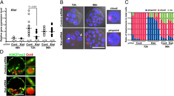
Transient repression of ectopic Xist in SCNT-generated embryos by Xist-siRNA injection. (A) Quantitative RT-PCR of Xist in cloned embryos injected with control or Xist-siRNA and cultured for 48 h (four-cell), 72 h (morula), or 96 h (blastocyst). The expression levels of Xist were significantly decreased by Xist-siRNA at 72 h (P < 0.001 by Student's t test), whereas no significant difference was observed at 48 or 96 h. (B) RNA-FISH analyses of Xist in siRNA-injected cloned embryos. After 72 h in culture (morula), ectopic Xist expression was observed as a cloud pattern in most nuclei of the control cloned embryos, whereas it was detected as small pinpoint signals in Xist-siRNA–treated cloned embryos (arrows). At 96 h (blastocyst), the majority of nuclei showed cloud signals of Xist RNA in both groups. (Scale bar, 50 μm.) (C) The ratios of blastomeres classified according to the cloud or pinpoint expression patterns of Xist analyzed by RNA FISH. Each column represents a single embryo. It is apparent that the siRNA against Xist strongly repressed spreading of the Xist RNA over the X chromosome at 72 h but not at 96 h. (D) Immunostaining for H3K27me3 (green) and Oct4 (red) in control or Xist-siRNA–treated cloned embryos at 96 h. Strong punctate signals of H3K27me3 were observed in more than 65% of trophectoderm cells (Oct4-negative; Upper Inset) and inner cell mass cells (Oct4-positive; Lower Inset) in both siRNA-treated groups. (Scale bar, 50 μm.)

