Fig. 3.
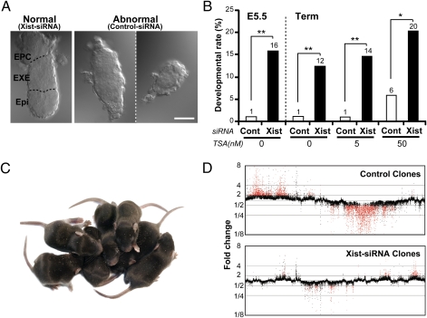
Effects of Xist-siRNA on the postimplantation development of cloned embryos. (A) Representative photomicrographs of siRNA-treated cloned embryos recovered at E5.5. Most Xist-siRNA embryos showed normal morphology (Left) with a distinct embryonic epiblast region (Epi) and extraembryonic ectoderm (EXE) or an ectoplacental cone (EPC), whereas most control siRNA-treated embryos showed abnormal morphology such as developmental retardation or ambiguous embryonic and extraembryonic regions (Center and Right). (Scale bar, 50 μm.) (B) The developmental rate of embryos assessed at E5.5 and at term. In some experiments, TSA was added at 5 or 50 nM in the culture medium. The numbers at the top of the bars indicate the rates of normal-shaped embryos (E5.5) and full-term births per embryos transferred. *P < 0.05, **P < 0.001 by Fisher's exact test. (C) A litter of cloned pups produced by SCNT from Sertoli cell nuclei, obtained following treatment with Xist-siRNA and 50 nM TSA. They were born at the best efficiency we observed in this series: Seven pups were born from 23 embryos transferred (30%) to a single recipient mother. (D) Gene expression profiles of livers in neonatal mice generated by SCNT with or without Xist-siRNA injection. The values indicate the mean fold changes from the control IVF level (=1). Red dots represent genes of which expression levels exceeded a twofold change in all of the individual clones. Xist-siRNA clones showed much fewer dysregulated genes compared with control clones. For data on each individual clone, see Fig. S2.

