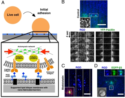
Fig. 1.
Formation of contractile pair assembly during initial adhesion. (A) Experimental schematic of live cells over nano-patterned supported lipid bilayer membranes. RGD peptides were chemically linked to lipids in supported bilayer membranes, and then bound to integrin receptors on the plasma membrane. (B) Contractile pair assembly of ligated RGD-integrin clusters across nano-patterned metal lines during initial adhesion. RGD-integrin clusters and associated YFP-paxillin laterally moved towards each other and piled up against the physical barrier of metal lines (two right boxes, 2 μm gap spacing between lines). (C) Preincubation with 50 μM blebbistatin blocked contractile movement of RGD-integrin clusters. White trajectories over each RGD cluster were 90 s time-projection of cluster positions. Inset overlay: Lifeact-Ruby (red) and EGFP-myosin light chain (green). (D) Colocalization of RGD and EGFP-integrin-β3 during early cell adhesion. Inset overlay: bright-field image. Membrane-bound RGD peptides attached to Cascade Blue neutravidin were utilized as reporters for activated integrins. (Scale bars, 5 μm).

