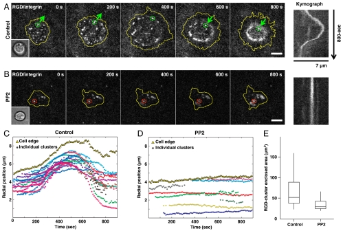
Fig. 4.
Outward movement of integrin clusters follows protrusion of cell edges. (A) Long-ranged outward then inward translocation of ligated integrin complexes. (B) Preincubation of 10 μM PP2, a Src-kinase inhibitor, blocked outward movement and cell spreading. Inset overlay: bright-field images, respectively. Kymographs: lateral movement of marked clusters, respectively. (C, D) Radial positions of each cluster’s trajectory (colored aster) and averaged cell edges (yellow triangle) of (A, 27 clusters) and (B, 8 clusters). (E) Cell contact area under control condition and PP2 treatment. Boxes, 1st and 3rd quartiles; whiskers, 10th and 90th percentiles; total 50 cells. (Scale bars, 5 μm).

