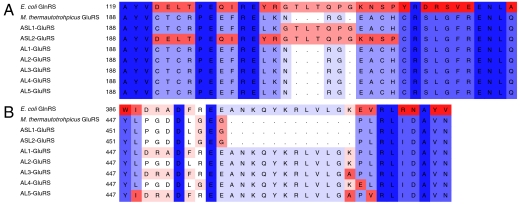
Fig. 4.
Alignment of E. coli GluRS, M. thermautotrophicus GluRS (WT-GluRS), and GluRS variants. Regions surrounding the (A) acceptor stem loop and the (B) anticodon loop are shown. GluRS variants only differ from WT-GluRS at positions indicated. The enzymes are otherwise sequence identical. Sequences are color coded according to amino acid identity (descending from blue to red).

