Fig. 1.
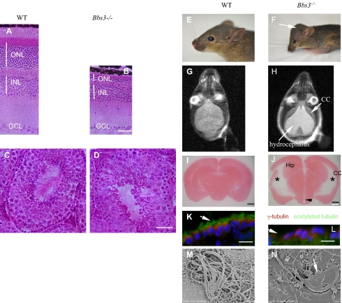
Photoreceptor cell loss, absent sperm flagella, and severe hydrocephalus in Bbs3−/− mice. H&E-stained WT (A) and mutant (B) eyes show loss of photoreceptor inner and outer segments, as well as degeneration of the outer nuclear layer. Scale bar, 100 μm. H&E-stained 4-mo-old WT (C) and Bbs3−/− (D) seminiferous tubules show a lack of spermatozoa flagella. Scale bar, 100 μm. (E and F) Pictures of 3-wk-old WT and Bbs3−/− brains. Arrow points to the domed cranium in a Bbs3−/− mouse. (G and H) MRI images of WT and Bbs3−/− brains. The long arrow indicates a hydrocephalic region and the short arrow points to thinning of the cerebral cortex. (I and J) Neutral-red–stained 60- to 100-μm thick coronal brain sections from WT (I) and mutant mice (J) show enlarged lateral ventricles (*), an enlarged dorsal third ventricle (arrowhead), reduced hippocampus (Hip), and thinning of the cerebral cortex (CC). Scale bar, 1 mm. Immunofluorescent staining of brain ventricle ependymal cells using antiacetylated tubulin (green) and anti–γ-tubulin antibody (red) reveals the lack of cilia and shortened cilia (arrow) in the ependymal cells of Bbs3−/− mice (L) compared to WT (K). Scale bar, 20 μm. (M and N) Scanning electron microscopy showed cilia morphological abnormalities in cultured ependymal cells from Bbs3−/− mice. Shortened and paddle-shaped cilia (arrow) are seen in Bbs3−/− ependymal cell cultures.

