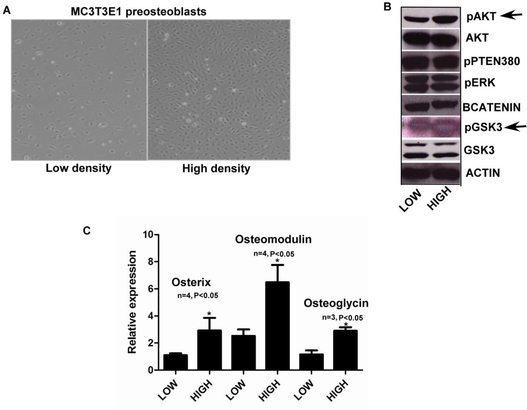
FIG. 2). Activation of Osterix, Osteomodulin and Osteoglycin in a P13K dependent manner.
Fig. 2A) MC3T3E1 cells plated at low and high density were then used for assaying gene expression using real time PCR and Western blotting for protein levels. Fig. 2B) When cells plated at low and high density were assayed for pAKT and total AKT levels using western blots we saw an increase in active AKT levels at high density along with increase in pGSK3S9 (pointed out with black arrows). There was no change in pERK or pPTEN levels (all the western blot images shown are representative of at least two different experiments). Fig. 2C) MC3T3E1 cells plated at sub confluent (low density) and confluent densities (high density), when assayed for osteoblast gene markers showed increases in Osterix, Osteomodulin and Osteoglycin gene expression using real time PCR. (n=3, p<0.05, all error bars indicate SD)

