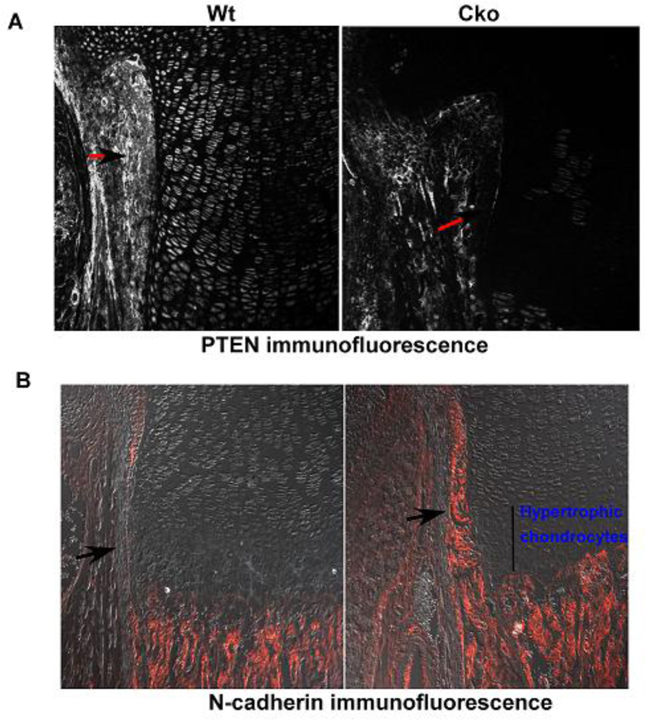
FIG. 6). Effect of loss of PTEN on adherens junctions.
Fig. 6A) Wild type and Pten cko new born tibial sections, PTEN has been deleted with Dermo1cre, showing PTEN protein levels using immunfluorescence, arrows with black heads and red tails point out the cells in the perichondrium that have Pten protein expression in the wildtype and its loss in the cko Fig 6B) N-cadherin immunofluorescence in the Pten cko the perichondrium shows increased N-cadherin expression compared to the wildtype control, the panels show N-cadherin expression in the cells of the perichondrium (pointed out with black arrows) showing N-cadherin positive cells.

