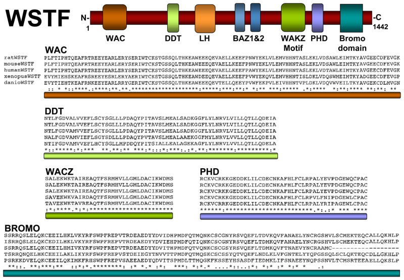
Figure 1.
Conserved motifs in WSTF. Top: schematic diagram of WSTF depicting the relative location of conserved protein domains LH, WAC, DDT, BAZ1, BAZ2, WAKZ, PHD and bromodomain. Bottom: sequence alignments of the specified domains illustrate the high degree of conservation among multiple species. “*” the residues or nucleotides in that column are identical in all sequences in the alignment; “: “ conserved substitutions have been observed; “.” semi-conserved substitutions are observed.

