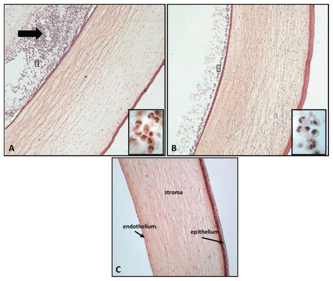
Figure 3.
Ocular histology from representative eyes infected with either the (A) parent, (B) the mutant strain, or (C) uninfected control. Boxed inserts (A & B) show higher magnification of PMNs in the anterior chambers of infected eyes. The corneas of the rabbits infected with the parent strain showed an influx of cells at the endothelium and in the anterior chamber (black arrow). Original magnification, 40X. Boxed insert magnification, 1000X.

