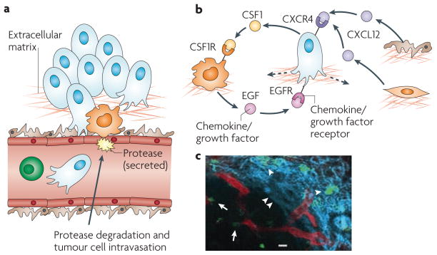
Figure 2. The invasive microenvironment.
a | Cancer cell intravasation into the blood circulation preferentially occurs in close proximity to perivascular macrophages. Disruption of endothelial cell contacts and degradation of the vascular basement membrane is required for cancer cell intravasation, which is mediated by proteases supplied from the cancer cells, macrophages or both. b | Cancer cell migration is controlled through a paracrine loop involving colony-stimulating factor 1 (CSF1), epidermal growth factor (EGF) and their receptors, which are differentially expressed on carcinoma cells and macrophages, resulting in movement of cancer cells towards macrophages (dashed arrow). Additional paracrine loops exist between cancer cells expressing C-X-C chemokine receptor 4 (CXCR4) and stromal cells, such as fibroblasts and pericytes, producing the cognate ligand stromal cell-derived factor 1 (SDF1, also known as CXCL12), which contribute to directional cancer cell migration. c | Tumour-associated macrophages (green) can be visualized in mammary tumours in living animals, in proximity to blood vessels (red), as indicated by arrows, and migrating along collagen fibres (blue, visualized by second harmonic resonance) as indicated by arrowheads. Part c reproduced, with permission, from REF. 54 © (2007) American Association for Cancer Research.

