Fig. 2.
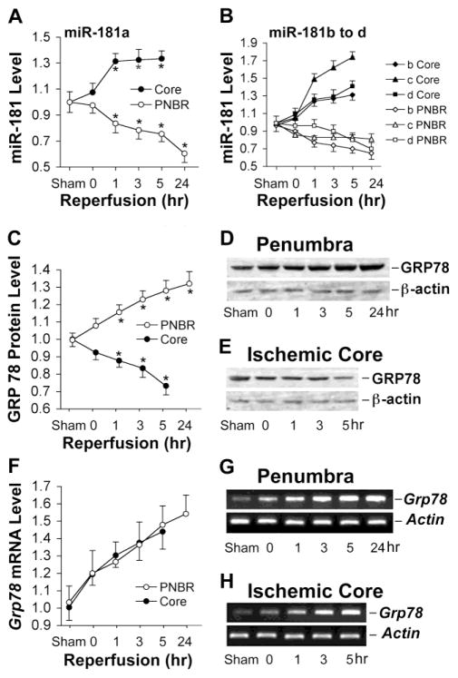
Expression of miR-181, GRP78 protein, and Grp78 mRNA after transient focal ischemia (MCAO). A. miR-181a expression in ischemic core and penumbra at different durations of reperfusion after 1 hr MCAO in mice shows increased levels in core but decreased levels in the penumbra (PNBR). B. miR-181b, c, and d show similar changes after ischemia, increasing in the core and decreasing in the penumbra. C. GRP78 protein decreases in the ischemic core and increases in the penumbra with increasing durations of reperfusion after MCAO. Quantitation by densitometry of westerns for 4 mice at each timepoint. D, E. Examples of western blots for GRP78 isolated from penumbra and ischemic core with increasing reperfusion time. F. Expression of Grp78 mRNA in ischemic core and penumbra increases with reperfusion time after MCAO. G, H. Examples of RT-PCR products from RNA harvested at increasing times after MCAO. Actin was used as the loading control for westerns and internal control for RT-PCR. N=4 mice/group in all experiments. *P<0.05 by ANOVA and Newman-Keuls post hoc test.

