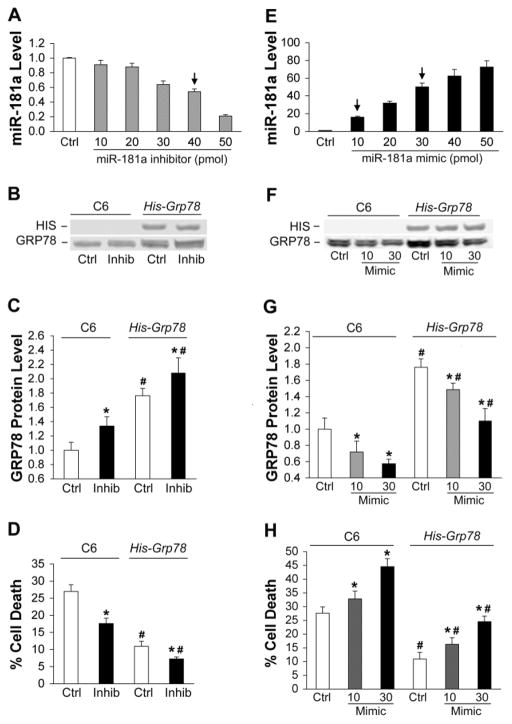
Fig. 4.
Effect of miR-181a mimic and inhibitor on C6 glia with or without GRP78 overexpression. A. Dose-response of miR-181a levels to transfection with increasing amounts of miR-181a inhibitor in C6 cultures. N=3. The arrow indicates the concentration used for B, C, and D of this figure. B. Representative blot shows miR-181a inhibitor increases GRP78 protein expression both in parental C6 cells and in the C6 line that stably overexpresses His-Grp78 under the control of the CMV promoter and lacking the native 3′UTR. C. Bar graph showing quantitation of significant changes in GRP78 protein expression with miR-181a inhibitor. D. miR-181a inhibitor reduces injury induced by 8 hr glucose deprivation (GD) in C6 cultures and His-Grp78 overexpressing cultures. E. Dose-response of miR-181a levels after transfection with increasing amounts of mimic in C6 cultures. N=3. The arrows indicate the concentrations used for F, G, and H of this figure. F. Representative blot shows miR-181a mimic decreases GRP78 protein expression in both C6 and C6- His-Grp78 overexpressing cells. G. Bar graph shows quantitation of GRP78 protein expression after transfection with miR-181a mimic. Protein levels in Ctrl C6 cells and His-Grp78 overexpressing cells with 30 pmol mimic were not statistically different. N=3. H. miR-181a mimic aggravates injury induced by 8 hr GD in both C6 and His-GRP78 overexpressing C6 cells. Cell death in Ctrl C6 cells and His-Grp78 overexpressing cells with 30 pmol mimic were not statistically different. All experiments were performed 3 times in triplicate. *P<0.01 compared with same cell line control (Ctrl) and #P<0.01 compared with the same dose of miR-181 inhibitor or mimic in C6 group by ANOVA and Newman-Keuls post hoc test.

