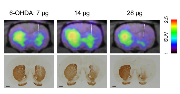
Figure 2.
Comparison between [18F]FDOPA-PET images and TH immunostaining. Upper panels show summated coronal PET images (from 11 to 90 min) of representative rats injected with 7 μg (left), 14 μg (middle), and 28 μg (right) of 6-OHDA. Lower panels show photomicrographs of TH-immunostained sections corresponding to PET images from the same rats. Scale bar = 100 μm.

