Fig. 2.
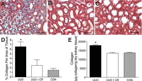
Mast cell stabilization reduces tubulointerstitial fibrosis in UUO kidney. A: representative section of 14-day UUO rat kidney stained with trichrome. Fibrosis is seen by the extensive collagen staining (blue). Scale bar = 50 μm. B: representative section of 14-day UUO kidney from rat treated with cromolyn sodium (CR) stained with trichrome. Note the absence of collagen staining. Scale bar = 50 μm. C: representative section of trichrome-stained 14-day kidney from CON rat. Scale bar = 50 μm. D: graph of mean percentage of collagen (±SE) per area of tissue as analyzed by spectral separation in trichrome-stained kidney sections, collagen being represented by blue-stained areas of tissue. UUO kidney (n = 5) displayed significantly more collagen staining (P < 0.05) than either the UUO kidney from rats treated with the mast cell stabilizer (UUO+CR; n = 3) or the kidney analyzed from CON rats (n = 4). E: graph of pepsin-soluble collagen content measured by biochemical assay in homogenates from rat UUO kidneys (14 day, n = 3), 14-day UUO kidney from rats treated with cromolyn sodium (UUO + CR; n = 3), and kidney from CON rats (n = 4). Values are means ± SE. The pepsin-soluble collagen content was significantly greater in UUO kidney homogenate compared with homogenate from UUO+CR or CON rats (*P < 0.05).

