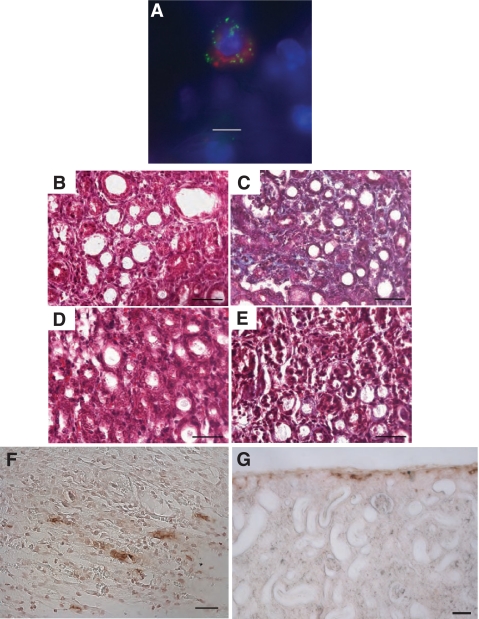
Fig. 5.
Losartan treatment curtails renal fibrosis in UUO kidney from CC mice. A: mast cells in UUO kidneys from CC mice immunoexpress renin. Shown is a representative section of fixed 14-day UUO kidney from CC mouse showing an avidin-labeled mast cell [conjugated to rhodamine (red)] costained with a polyclonal anti-renin antibody (green). The staining is not colocalized. B: representative section of Masson's trichrome-stained 14-day UUO kidney from a CC mouse treated with losartan. Note the lack of collagen staining (blue). Scale bar = 50 μm. C: representative section of Masson's trichrome-stained 14-day UUO kidney from a CC mouse. Collagen is stained blue. Scale bar = 50 μm. D: representative section of Masson's trichrome-stained 14-day UUO kidney from a MCD mouse treated with losartan. Scale bar = 50 μm. E: representative section of Masson's trichrome-stained 14-day UUO kidney from a MCD mouse. Scale bar = 50 μm. F: representative section of UUO kidney from a CC mouse stained with an anti-chymase antibody using an immunoperoxidase technique. Chymase-positive mast cells were frequently observed in medulla. Scale bar = 20 μm. G: representative section of UUO kidney from a CC mouse treated with losartan and stained with an anti-chymase antibody. Scale bar = 50 μm.

