Fig. 7.
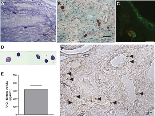
Human kidney mast cells express active renin. A: fixed section of archived sample of fibrotic human kidney stained with toluidine blue. The mast cells are peritubular. Scale bar = 10 μm. B: fixed section of archived sample of fibrotic human kidney costained with avidin-HRP for identifying mast cells (brown) and Gomori's trichrome for fibrotic regions (blue). Scale bar = 25 μm. C: fixed section of archived fibrotic human kidney showing a mast cell costained with goat anti-renin antibody conjugated to Alexa Fluor 594 (red) and avidin-FITC (green). Scale bar = 10 μm. D: mast cells immunomagnetically isolated from normal kidney and stained with toluidine blue. Scale bar = 10 μm. E: measurement of ANG I-forming activity (in pg·ml−1·h−1) in the lysate from isolated human kidney mast cells (±SE; n = 3). F: fixed section of archived fibrotic human kidney stained with an anti-chymase antibody using an immunoperoxidase technique. Scale bar = 50 μm.

