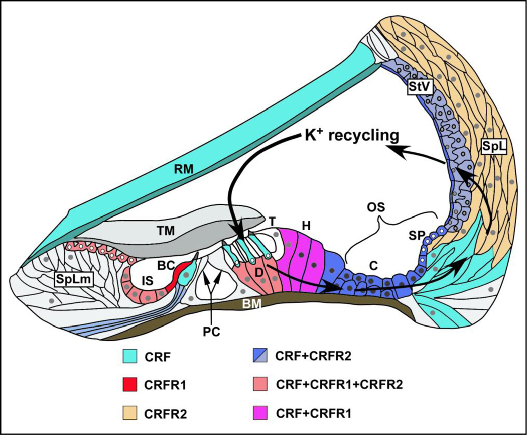
Figure 3. Schematic CRF, CRFR1 and CRFR2 of expression in the cochlea in relation to routes of potassium recycling.
CRF (teal) is expressed in inner hair cells (IHC) and outer hair cells (OHC). CRF is co-expressed with CRFR2 in afferent spiral ganglion neurons (only fibers shown here) and in the stria vascularis (StV, shown in shades of blue, reflecting level of expression; dark blue- high expression, lighter blue- lower expression). CRF is co-expressed with CRFR1 and CRFR2 in Deiter’s cells (D) below outer hair cells (orange), while in Hensen’s cells (H), lateral to the Deiter’s cells, CRF is co-expressed with CRFR1 (pink). In the support cell populations not immediately adjacent to hair cells, CRF is co-expressed with CRFR1 and CRFR2 in the inner sulcus (IS) and in interdental cells (orange) of the Spiral Limbus (SpLm), while in support cells lateral to the organ of Corti (blue), such as Claudius cells (C) Boettcher cells (below Claudius cells), and cells of the outer sulcus such as those lining the spiral prominence, CRF is co-expressed only with CRFR2. CRFR1 (red) is expressed alone (without CRF) in the border cell (BC) adjacent to the medial surface of the IHC. (BC-Border cell; BM- basilar membrane; C- Claudius cells; D- Deiter’s cells; H- Hensen’s cells; IS- inner sulcus; OS- outer sulcus; PC- Pillar cells; RM- Reissner’s membrane; SP- spiral prominence; SpL- spiral ligament (containing fibrocytes populations); SpLm- spiral limbus; StV- stria vascularis; T- Tectal cell; TM- tectorial membrane; Figure adapted from Jentsch, 2000.)

