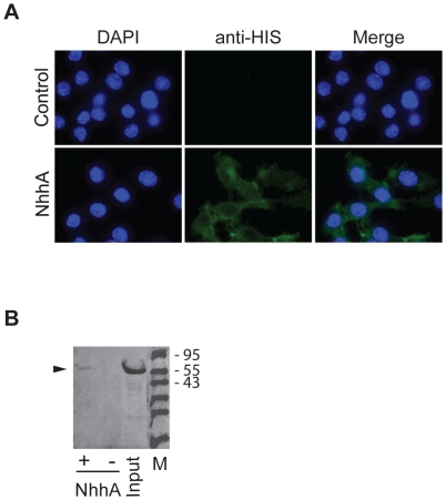
Figure 1. NhhA binds to macrophages.
A: The RAW 264.7 mouse macrophage cell line was grown on coverslips and incubated with 400 nM recombinant NhhA for 4 hours. Immunofluorescence analysis was performed using a monoclonal mouse anti His-tag antibody followed by an Alexa Fluor 488-labeled anti-mouse antibody. Control cells were incubated with an unrelated His-tagged protein (NMC0101). One representative experiment of three is shown. B: Macrophages were incubated with 400 nM recombinant NhhA (+ NhhA) or NMC0101, an unrelated His-tagged protein (− NhhA) for 4 hours. Cells were washed in PBS, dissolved in SDS-PAGE sample buffer and cell-bound material or input material was analysed by immunoblot analysis using an anti His-tag antibody. Arrowhead indicates migration of NhhA. The sizes in kDa of a protein ladder is indicated. One representative experiment of three is shown.

