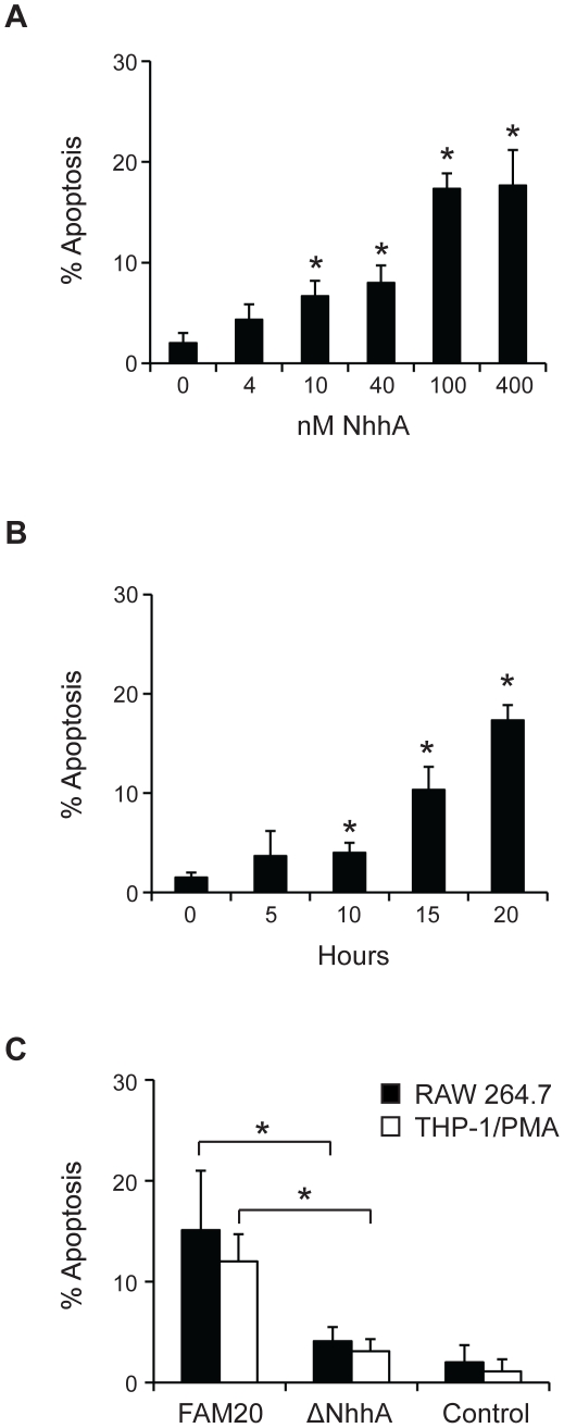
Figure 3. Effect of recombinant and endogenous NhhA on macrophage apoptosis.
A: The RAW 264.7 mouse macrophage cell line was incubated with 0–400 nM recombinant NhhA for 20 hours. B: RAW 264.7 cells were incubated with 400 nM NhhA for 0–20 hours. C: RAW 264.7 or PMA-differentiated THP-1 cells were infected with the N. meningitidis serogroup C strain FAM20 or a NhhA-deficient mutant of FAM20 (ΔNhhA) at MOI = 100 for 20 hours. Control cells were uninfected. Apoptotic cells were visualized using the APOPercentage kit and the relative numbers of apoptotic cells were determined by light microscopy. Values indicate mean±SD of three independent experiments. *, p<0.05 versus untreated control(panel A+B) *, p<0.05 versus FAM20 treated cells (panel C).

