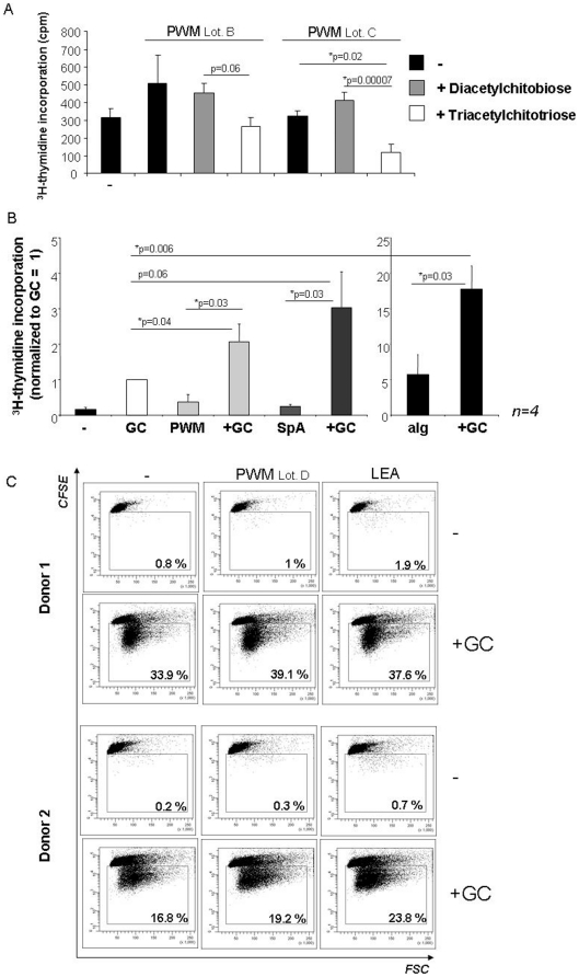
Figure 5. Contribution of the lectin component.
A: Human B cell proliferation in response to PWM (Lot. B and C) in the presence or absence of N,N-di-acetylchitobiose or N,N,N-tri-acetylchitotriose was assessed by 3H-thymidine incorporation given in counts per minute (cpm). The mean values ± SEM from one representative experiment performed in triplicates of n = 2 is shown. B: Human CD19+ B cells were left unstimulated or stimulated with 0.25 µM GpC PTO ODN (GC) and/or 10 µg/ml of PWM, 5 µg/ml SpA or 10 µg/ml anti-Ig (aIg) for 72 hours. Proliferation was quantified by 3H-thymidine incorporation. The diagram shows the means from n = 4 experiments ± SEM. The values obtained were normalized to GC = 1 (710±280 cpm = mean ± SEM). C: Human CD19+ B cells were stained with CFSE and stimulated with highly purified PWM (Lot. D) or Lycopersicon esculentum lectin (LEA) with or without GpC PTO ODN (GC). Proliferation was quantified by CFSE dilution on day 4. The percentage of proliferating cells is provided in each dot plot. The results from two independent donors of n = 4 are shown.

