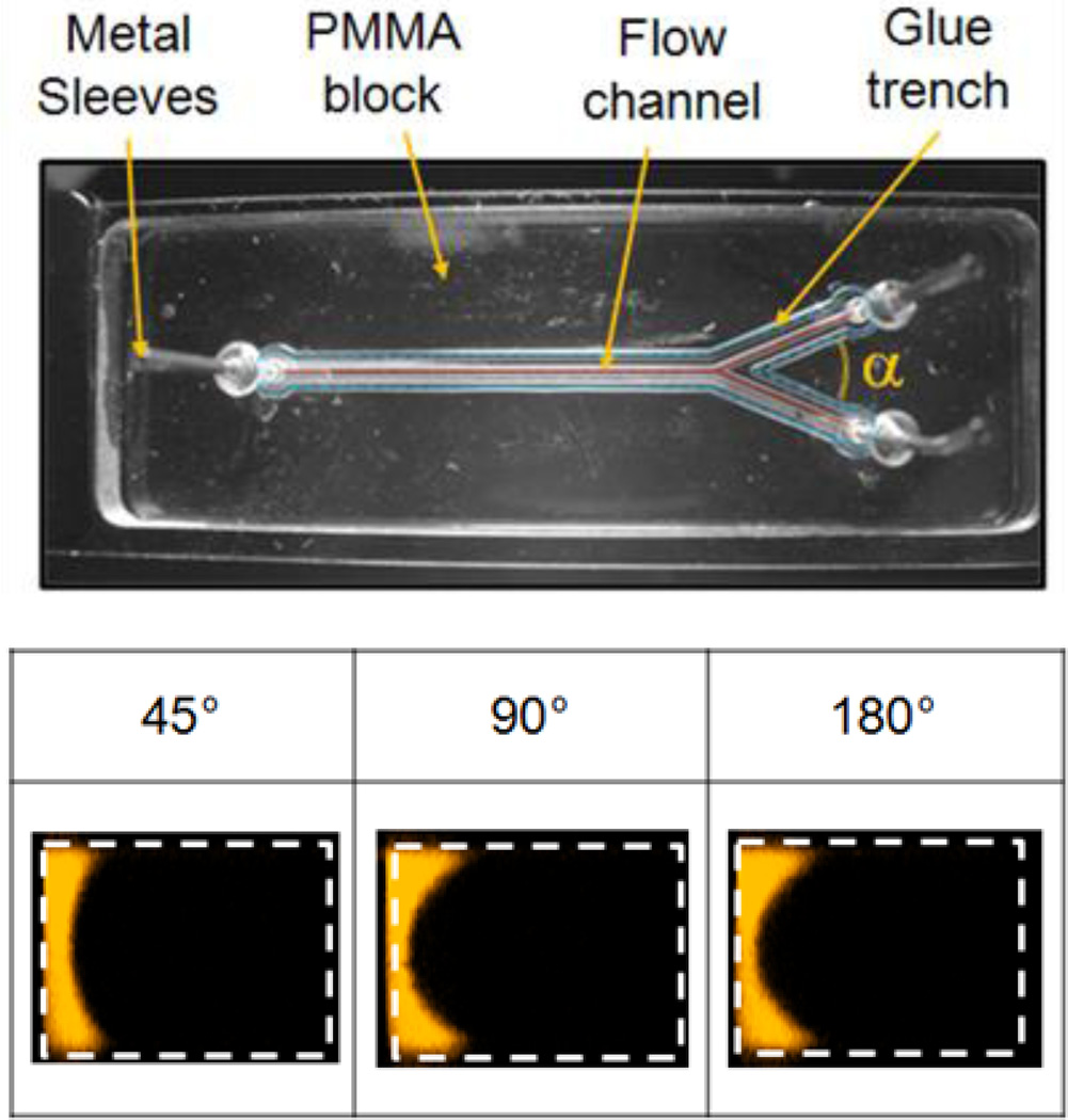
Figure 2.
Microchannels were fabricated from polymethylmethacrylate (PMMA) and attached to a glass slide using UV-curable glue. A trench around the boundary of the main channel prevented the glue from running into the channel. All channels were 600 µm wide and 400 µm deep (±10µm). Confocal cross-sectional images of the main channel show the focused region for three angles of confluence (α). Flow rates for the sheath and focused streams were 720 and 29 µL/min, respectively (Re ≈25). Deionized water was used for both streams with rhodamine dye added to the focused stream only [47]

