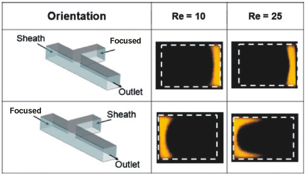
Figure 3.
Channel cross-sectional images from confocal microscopy show the concentration profiles for a conventional T-inlet channel design (α = 90°). The sheath and focused streams were switched for each case of the Re (10, 25). The first row shows the case in which the sheath stream was aligned with the main channel. The second row shows the results in which the focused stream was aligned with the main channel. The channel was 380 µm×600 µm (height×width). The flow rates for the focused stream and sheath flow, respectively, were 11 and 283 µL/min for Re ≈10 and 28 and 707 µL/min for Re ≈ 25 [9]

