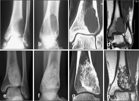
Fig. 2.
X-ray (a, b) and CT (c, d) images of a 24-year-old female patient diagnosed with an osteoclastoma of the distal tibia. The patient experienced pain when weight-bearing. TricOs bone substitute material was applied in combination with autologous cancellous bone from the iliac crest at a rate of 1:5. The defect had a volume of 10.5 cm3. At 18 months post surgery, CT scans were performed. The lesion was completely filled with newly formed tissue reminiscent of healthy cancellous bone. A sclerotic margin was observed. After 56 months the bone substitute material was radiographically still detectable. Resorption was determined as a stage 2 (e–g). Also, MRI was performed after 56 months to rule out recurrence of the osteoclastoma. A hypointensive zone was seen in the CT images corresponding to the sclerotic margin (h)

