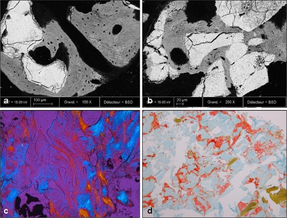
Fig. 5.
a, b Backscattered scanning electron microscopy (BSEM) demonstrating remnants of the transplanted bone substitute (white) material and new bone formation in grey and black. New bone formation is in close apposition to the biomaterial indicating good osseointegration. c Polarized light microscopy (10x) showed both residual granules and newly formed bone with haversian system. d In PMMA sections stained for Movat’s pentachrome (10x) new mineralized bone (green) and some unmineralized osteoid (red) as well as residual granules (light blue) were seen gradually being resorbed by osteoclastic cells

