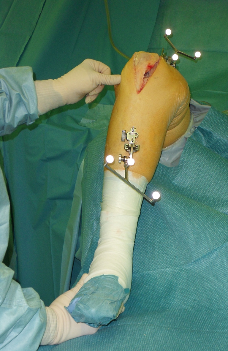
Fig. 2.

Measurement setup. The tensor is positioned in the joint gap; the femur is elevated by an electronic leg holder. The patella remains in the anatomical position; the surgeon controls the tibia to allow physiological knee rotation during knee flexion
