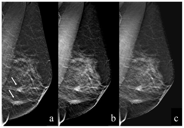
Figure 1.
MLO images of the left breast of a 59YO woman depicting 2 masses, both were pathology verified as IDC and DCIS. The actually ascertained FFDM (a), the synthetically reconstructed projection image (2D) from the 3D dataset (b), and one slice (1mm thick) from the tomosynthesis image set (c), are shown.

