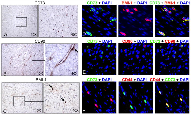
Figure 1. Immunohistochemical staining demonstrated stem cell marker expression in DTs.
Sixteen archived DTs were stained for stem cell markers CD73 (A), CD90 (B), and BMI-1 (C), and representative images demonstrated the presence of abundant MSCs (black arrows) within tumors (original magnification 10X or 40X). Fluorescent immunohistochemistry of paraffin-embedded DTs showed co-expression of the MSC markers CD73 (green) and BMI-1 (red) (D), CD73 (green) and CD90 (red) (E), as well as CD73 (green) and CD44 (red) (F). Co-expression of both markers produced a yellow overlay. Original magnification was 40X. In this and subsequent fluorescent immunohistochemistry, nuclei were counterstained blue with DAPI.

