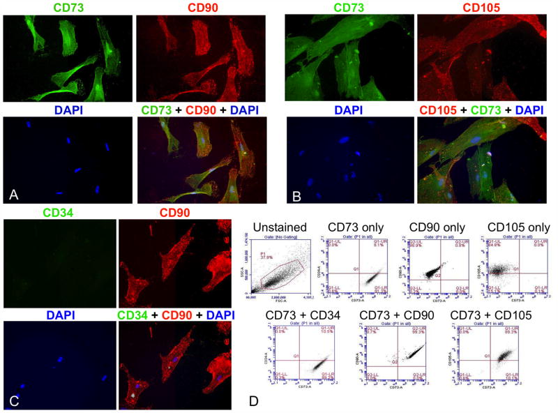
Figure 2. DT-derived MSCs co-expressed positive but not negative MSC markers.
Fluorescent immunohistochemistry was performed on early passage DT-derived MSCs demonstrating co-expression of the MSC markers CD73 (green) and CD90 (red) (A) as well as CD73 (green) and CD105 (red) (B), but not expression of the negative endothelial precursor marker, CD34 (C). Original magnification was 40X. FACS analysis of these cells showed co-expression of the obligate MSC markers CD73, CD90, and CD105, but not CD34 (D). The gated population is outlined in red on the top, left dot plot. Sorting showed that: 99.1% of cells were CD73+, CD34−; 99.3% were CD73+, CD90+; and 89.3% were CD73+, CD105+.

