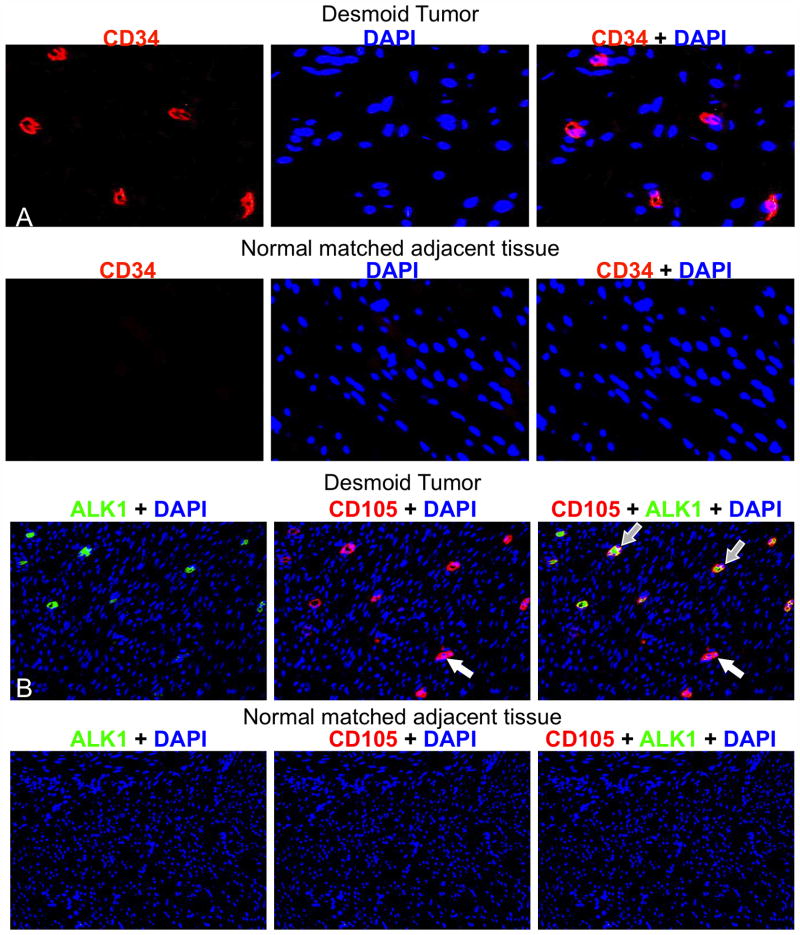
Figure 3. DTs contained CD34+, CD105+, and ALK1+ cells.
Representative fluorescent immunohistochemistry images are shown of formalin-fixed, paraffin-embedded DTs stained for CD34 (red) (A, Top). Matched normal adjacent tissue did not display any CD34+ cells (A, Bottom) (original magnification 40X). Representative fluorescent immunohistochemistry images of formalin-fixed paraffin-embedded archival DTs stained for ALK1 (red) and CD105 (green) (B, Top). Matched normal adjacent tissue showed only AKL1− and CD105− cells (B, Bottom). Open arrows indicate CD105+ ALK1− cells; grey filled arrows indicate dual positive cells. The original magnification was 20X.

