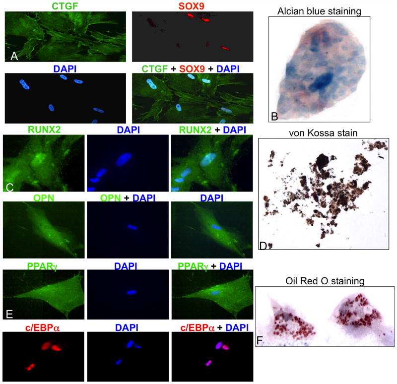
Figure 4. DT-derived MSCs exhibited tri-potent differentiation capability.
DT-derived cells were incubated in differentiation media, and confirmation of differentiation was obtained by expression of chondrocyte markers CTGF (green) and Sox9 (red), overlay shows yellow stain (A) plus positive Alcian Blue staining (B); osteocyte markers RUNX2 (green) and OPN (green) (C) plus positive von Kossa staining (D); and adipocyte markers PPARγ (green) and c/EBPα (red) (E) plus positive Oil Red O staining (F). Original magnification was 40X.

