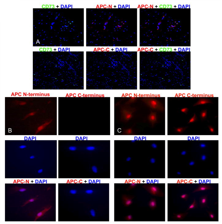
Figure 5. MSCs expressed only the APC-N terminus consistent with somatic APC+ LOH.
Fluorescent immunohistochemistry was performed on paraffin-embedded DTs and DT-derived cells for both the APC (N-15) and (C-20) termini. Normal human adipose-derived MSCs were used as controls. In MSCs from both the DT (A) and cell line (B), staining was positive for the N-terminus (red) but not the C-terminus. In contrast, staining was positive for both N- and C-termini of APC protein in normal human adipose-derived MSCs (C). Original magnification was 10X and 20X. Overlay shows a purple nuclear stain.

