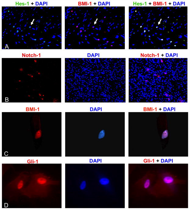
Figure 7. Notch and Hedgehog pathways were upregulated in DTs.
Fluorescent immunohistochemistry was performed on paraffin-embedded DTs and showed positive expression of the Notch target gene Hes-1 (green) (A) and Notch-1 (red) (B). Co-localization with BMI-1 (red) produced yellow nuclei (A). DT-derived cells also expressed the transcriptional repressor BMI-1 (C). Co-localization of red staining of BMI-1 with the blue counter-stain produced purple-colored nuclei. Similarly, purple nuclei were evident when the MSCs were stained with antibodies for the Hedgehog transcriptional activator, Gli-1 (red) (D).

