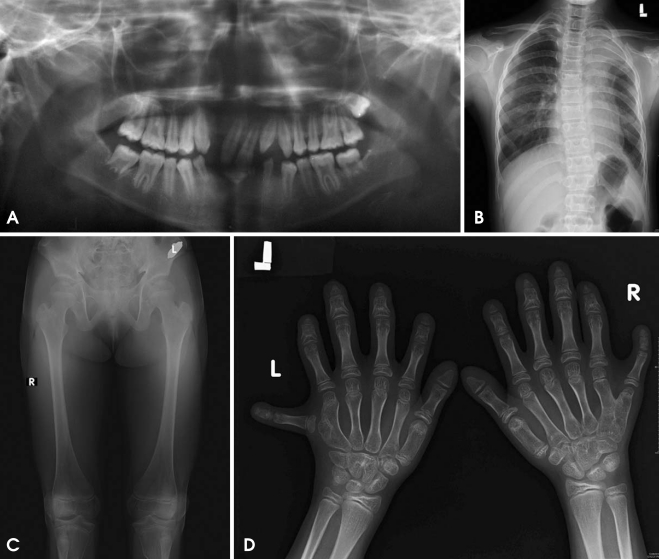
Fig. 3.
A. Panoramic radiograph shows conical shaped teeth, missing mandibular permanent anterior, retained deciduous mandibular canine and right lateral incisor. B. Chest radiograph shows homogenous opacity in the left upper zone. C. Antero-posterior view of legs shows Genu valgum. D. Hand-wrist radiograph shows carpal fusion, postaxial polydactyly, shortening of metacarpal and phalangeal bone with cone shaped epiphysis and fusion of capitate and hamate on right hand and hamate and triquadral on left hand.

