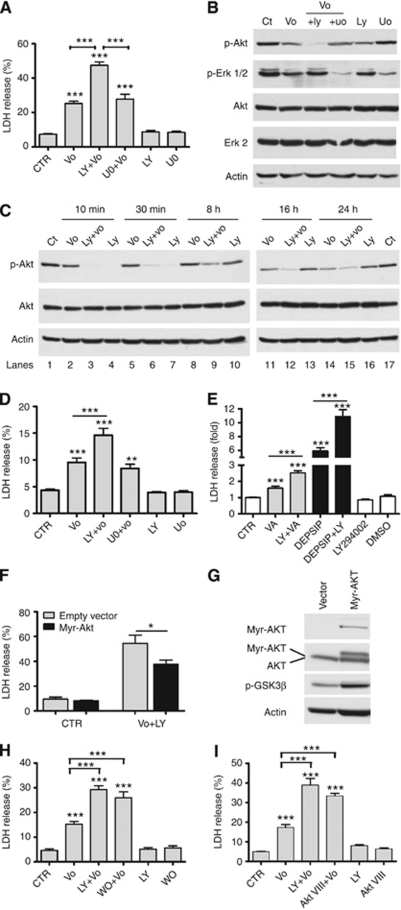
Figure 2.
PI3K-AKT inhibitors enhancement of vorinostat (vo)-induced cytotoxicity is mediated by strong and durable AKT inhibition. (A) LDH viability assay for SCC25 cells treated with vehicle (CTR) or a maximal cytocidal dose of vo (5 μM) alone or in combination with LY (10 μM) or uo (10 μM) for 24 h. (B, C) Western blots show lysates from SCC25 cells treated with vo (5 μM) alone or in combination with LY (10 μM) or uo (10 μM) at distinct time points. (D) LDH viability assay for Cal27 cells treated with vehicle (CTR) or vo (5 μM) alone or in combination with LY (10 μM) or uo (10 μM) for 24 h. (E) LDH viability assay for SCC25 cells treated with valproic acid (VA, 3 mM) or Depsipeptide (Depsip, 5 nM) alone or in combination with LY (10 μM). (F) SCC25 cells were transfected with constitutively active AKT (myr-AKT, black boxes) or its corresponding empty vector (open boxes) and then left untreated (CTR) or were treated for 24 h with vo (5 μM)+LY294002 (10 μM, LY). Cell viability was then estimated by LDH release. (G) Western blot of lysates from SCC25 cells showing the expression of the myr-AKT and endogenous AKT in vector only and myr-AKT transfected cells. Phosphorylation of the AKT target GSK3β (p-GSK3β) are provided to confirm functional AKT activity. Western blot figures are representative of two independent experiments. (H) LDH viability assay for SCC25 cells treated with vo (5 μM) alone or in combination with LY (10 μM) or wortmannin (1 μM). (I) Similar assay for cells treated with vo (5 μM) alone or in combination with AKTVIII (10 μM) for 24 h. LDH values presented as a percent of total LDH. Western blot figures are representative of at least three independent experiments. *Indicates P⩽0.05. **Indicates P⩽0.01. ***Indicates P⩽0.001 vs CTR. Values presented as mean±s.e.m. of at least three independent experiments performed in triplicate.

