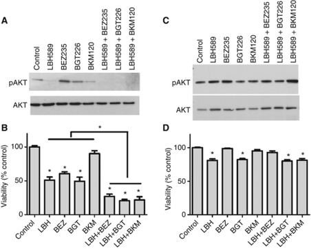
Figure 3.
Clinically relevant PI3K-AKT-mTOR inhibitors enhance cancer cell specific cytotoxicity induced by LBH589. (A) Western blots show lysates from SCC25 cells treated for 48 h with LBH589 (300 nM), BEZ235 (300 nM), BKM120 (300 nM) and BGT226 (300 nM), alone in combinations. (B) Viability assay for SCC25 cells subjected to the same treatments as in (A). (C) Western blots show lysates from normal HKs subjected to the same treatments as in (A). (D) Viability assay for normal HKs cells subjected to the same treatments as in (C). Western blot figures are representative of three independent experiments. Values are means±s.e. of three independent experiments performed in triplicate. *Indicates P<0.05.

