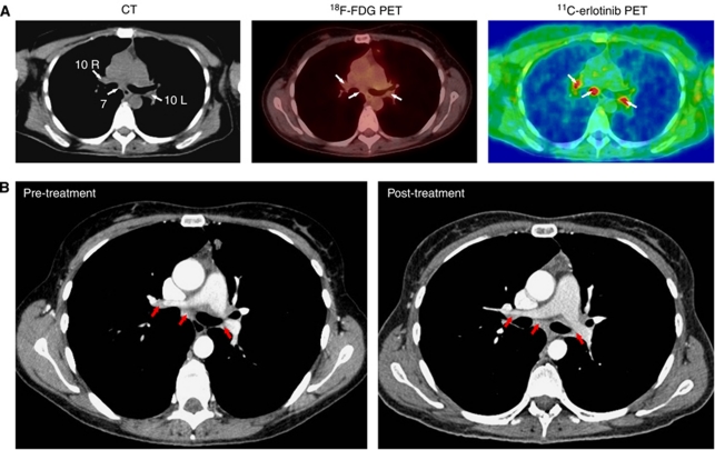
Figure 2.
11C-erlotinib accumulation in lymph nodes that were negative on 18F-FDG PET/CT in a 42-year-old patient (no. 6). (A) Left: transaxial slices of contrast-enhanced CT; middle: 18F-FDG PET/low-dose CT; right: 11C-erlotinib PET/low-dose CT. CT (left figure) showed an enlarged lymph node (>10 mm) at position 7 (arrow) and non-enlarged lymph nodes (<10 mm) at positions 10R and 10L (arrows). None of these lymph nodes were visualized by 18F-FDG PET/CT (arrows, middle figure), whereas both enlarged and non-enlarged lymph nodes were visualized by 11C-erlotinib PET/CT (arrows, right figure). The ratio between 11C-erlotinib average radioactivity concentrations in the lymph nodes and that in surrounding lung tissue was 2. (B) Comparison of pre-treatment and 1-year post-treatment CT scans showed no significant change in the size of any of these lymph nodes.

