Figure 3.
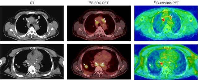
11C-erlotinib PET/CT demonstrates the heterogeneous nature of advanced lung cancer. Two transaxial slices of (left) contrast-enhanced CT; (middle) 18F-FDG PET/low-dose CT, and (right) 11C-erlotinib PET/low-dose CT. A 48-year-old patient (no. 7) with NSCLC in the right lung and enlarged mediastinal lymph nodes (upper and lower panel left figure, arrows). Both 18F-FDG and 11C-erlotinib accumulated in lymph nodes at positions 4R and 5 (upper panel, arrows). The tumour in the right lung and one of the lymph nodes at position 7 showed only a weak accumulation of 11C-erlotinib (arrows). The ratio between the 11C-erlotinib average radioactivity concentrations in the lymph node metastasis and that in surrounding lung tissue was 2 (see Figure 4).

