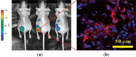
Figure 3.
(a) Left panel: In vivo fluorescence imaging of three nude mice bearing MCF-7/HER2 xenografts implanted in the lower back 30 h after i.v. injection with anti-HER2 QD-ILs; (b) Right panel: A 5 μm section cut from frozen tumor tissues harvested at 48 h postinjection and examined by confocal microscopy by a 63× oil immersion objective (image size, 146 μm × 146 μm). The tumor section was examined in two-color scanning mode for nuclei stained by DAPI (blue) and QD-ILs (red). (Cited from Weng et al. [107]).

