Fig. 6.
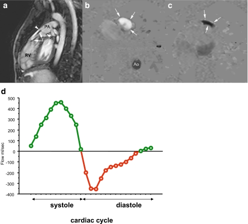
Severe pulmonary regurgitation post tetralogy of Fallot repair in a 20-year-old man. SSFP cine MRI (a) of the right ventricle (RV), RV out-flow tract and main pulmonary artery (PA) obtained at diastole. Abnormal coaptation of pulmonary cusps (arrowheads) results in severe pulmonary regurgitation (arrows). Phase image perpendicular to RV outflow tract during systole (b) indicates forward blood flow (“bright” appearance) across outflow tract during systole (arrows). Corresponding phase image during diastole (c) shows severe backward flow (“black” appearance), resulting in regurgitation fraction of 46%, and reduced RV ejection fraction of 43% (d forward flow is shown in green, severe regurgitant diastolic flow in red)

