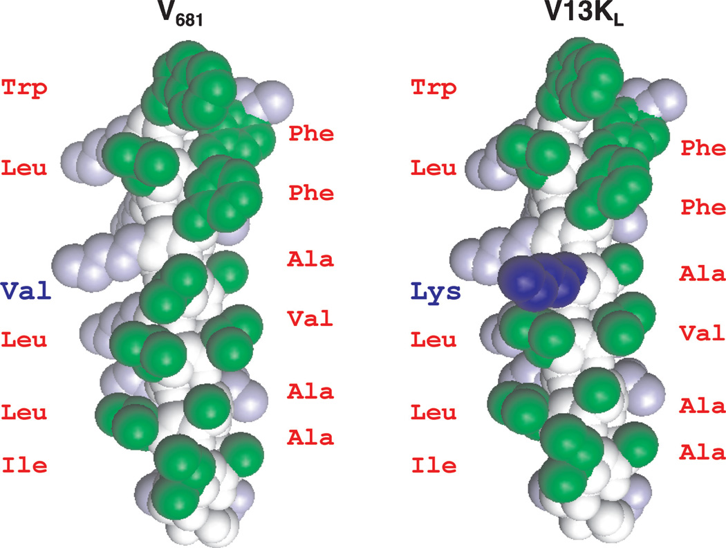
Figure 1. Space-filling model of peptides V681 and V13KL.
Hydrophobic amino acids on the non-polar face of the helix are green; hydrophilic amino acids on the polar face of the helix are gray; peptide backbone is colored white. The Lys substitution at position 13 (V13KL) on the non-polar face of the helix is blue. The models were created by the pymol v0.98. The peptide sequences are shown in Table 1.

