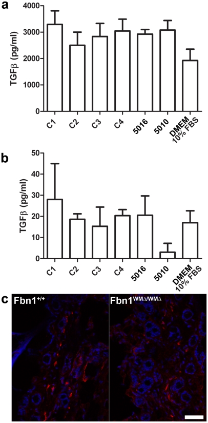
Figure 7. Measurements of TGF-β in cultured fibroblasts; α-smooth muscle actin staining.
(a) WMS fibroblasts (family members 5016 and 5010) secreted equal amounts of total TGF-β protein compared to controls (C1, C2, C3, and C4). (b) No significant differences were detected in amounts of active TGF-β present in the media of WMS fibroblasts compared to controls. Medium containing 10% fetal bovine serum was used to show baseline values. For experiments in (a) and (b), n = 2 or 3, and the error bars represent the standard deviation. (c) Skin from 6-month old wildtype and WMΔ/WMΔ littermates showed no difference in numbers of cells stained by an α-smooth muscle actin antibody (red). DAPI nuclear stain is blue. Scale bar = 20 µm.

