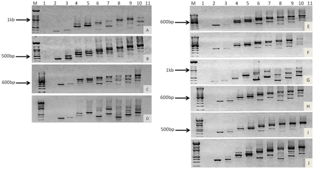
Figure 3. Amplification of CD44 variant exons.
RT-PCR was performed using exon specific primers (lanes 1–10 were exons v1 to v10, respectively), and resolved on a 1.5% agarose gel with a 100 bp ladder. NOK cells (A), DT cells (B), OSC-19 (C), and OSC-20 (D) and three matched primary and metastasis derived OPSCC cell line pairs; HN4 (E) and HN12 (F), HN22 (G) and HN8 (H), and HN30 (I) and HN31 (J) were assessed.

