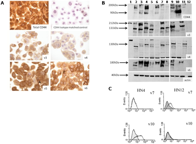
Figure 4. Detection of total CD44 and variant CD44 protein.
Representative figures of immunostaining with all cells stained positive in this field (A), western blotting (B), and flow cytometry (C). (A) Immunostaining of HN12 cells for total CD44, an isotype matched control, v3, v4, v5, and v6. (B) Western blotting lanes 1–12 contain MW marker, HaCaT, HN4, HN12, HN8, HN22, HN30, HN31, OSC-19, OSC-20, DT, and Namalwa cells, repectively. (C) flow cytometry results for HN4 and HN12 cells detecting v7 and v10.

