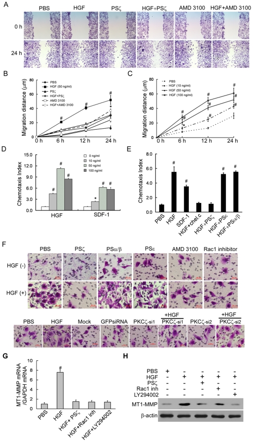
Figure 4. Overexpressed CXCR4 in HGF-stimulated MDA-MB-436 cells is functional.
(A–B). Confluent cells were grown in 0.5% FBS medium for 24 hours and were then wounded with a tip. The cells were washed, and the medium was replaced with or without addition of HGF, PSζ peptides or AMD3100. Representative micrographs of the wounds are shown together with the results of the migration quantification. Results are presented as the mean ± SD of 3 independent experiments. # P<0.01 as compared to PBS. Original magnification, 200×. (C).Dose and time-dependent response of HGF-induced MDA-MB-436 cell migration. (D).Dose-dependent response of HGF-induced MDA-MB-436 cell chemotaxis. (E).PKC and CXCR4 regulate HGF-mediated chemotaxis. MDA-MB-436 cells were incubated for 30 minutes with 10 µM chelerythrine chloride, an inhibitor of all PKC, or for 1 hour with 10 µM of PS-α/β, PS-ε, or PSζ peptide. (F). Boyden chamber assays were performed using the SDF-1 ligand for CXCR4 as a chemotactic attractive agent in the lower chamber. AMD3100, PSζ, PSα/β, PSε and NSC23766 (upper) or PKCζ-siRNA (lower) was added to the cell culture for the blocking assay. Data are shown as the mean ± SD of three experiments. A representative study is shown. (G). qRT-PCR analysis of the MT1-MMP mRNA extracted from MDA-MB-436 cells cultured for 24 hours with the indicated agents. Results are presented as the mean ± SD of three independent experiments. # P<0.01 as compared to PBS. (H). Western blot analysis of the total protein expression levels of MT1-MMP in MDA-MB-436 cells cultured for 24 hours with the indicated agents. The experiment was repeated three with similar results. A representative study is shown.

