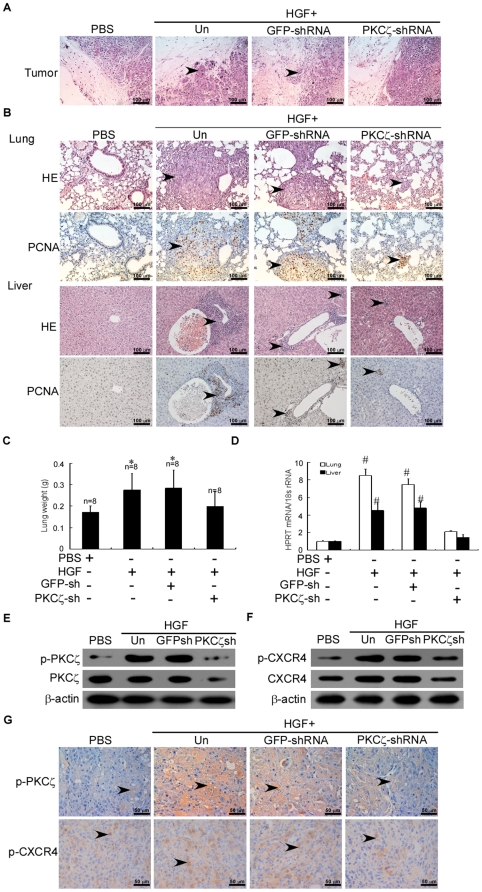
Figure 6. HGF enhances CXCR4 expression via PKCζ and promotes the invasion and metastasis of breast cancers in BALB/c-nu mice.
(A) Micrographs of H&E-stained xenografts demonstrating the presence or absence of margin invasion by the MDA-MB-436 tumors untransduced (Un) or transduced with GFP-shRNA or PKCζ-shRNA (original magnification, 200×). (B) Representative micrographs of lung (upper) or liver (lower) tissue sections with H&E staining and PCNA immunohistochemical staining (original magnification, 200×). Each group from two independent experiments. (C) Mean ± SD wet lung weight in tumor-bearing mice. The number of mice in each group is indicated. * P<0.05 as compared to PBS-treated mice. (D) Expression of human HPRT mRNA relative to mouse 18S rRNA as determined by qRT-PCR in the lungs and livers of tumor-bearing mice. Data were normalized to PBS-treated mice. # P<0.01 as compared to PBS-treated mice. (E–F) Immunoblot of total PKCζ and p-PKCζ or total CXCR4 and p-CXCR4 protein, respectively, in breast tumor xenografts in female nude mice that were inoculated in the mammary fat pads with MDA-MB-436 cells treated as indicated. The experiment was repeated twice with similar results. A representative study is shown. (G) Representative microphotos of immunohistochemical results for p-PKCζ (middle) or p-CXCR4 (lower) expression in MDA-MB-436 tumors (original magnification, 400×).

