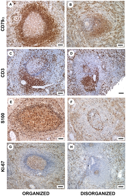
Figure 2. Leukocyte populations in organized and disorganized spleens.
Distribution of CD79α+ B and CD3+ T lymphocytes, S100+ dendritic cells and Ki-67+ proliferating cells in the spleens of dogs infected with L. infantum with and without disruption of splenic lymphoid tissue structure (figures A, B, C, D, G and H, bar = 70 µm; figures E and F, bar = 50 µm).

