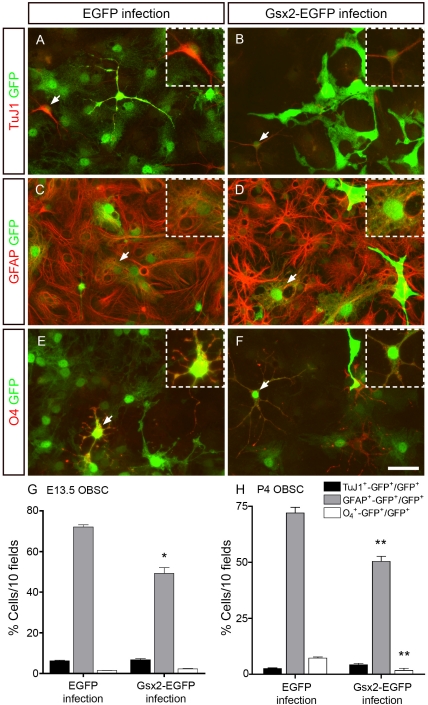
Figure 6. Transduction of Gsx2 decreases the capacity of OBSCs to differentiate into astrocytes and oligodendrocytes.
E13.5 OBSCs transduced with viral particles expressing EGFP and Gsx2-EGFP were plated under differentiation conditions for 6 (not shown) and 20 days (A–F). Dual immunostaining with GFP and TuJ1, GFAP or O4 antibodies revealed the populations of neurons (A, B) and oligodendrocytes (E, F) to be similar in cultures infected with either virus. However, GFAP+-GFP+ cells were 32% fewer in the Gsx2-EGFP infected cultures than in those infected with EGFP alone (C, D, G). The insets show high magnification images of the double-labeled cells indicated with arrowheads in the main images. The graph in G represents the mean percentage ± s.e.m. (n = 8 cultures per condition; data from 6 and 20 days of differentiation were combined). *P<0.05 versus the values obtained from EGFP-infected cultures. P4 OBSCs transduced with viral particles expressing EGFP and Gsx2-EGFP were plated under differentiation conditions for 6 days (H). Dual immunostaining with GFP and TuJ1, GFAP or O4 antibodies revealed 30% GFAP+-GFP+ cells and 4-fold O4 +-GFP+ fewer cells in the Gsx2-EGFP infected cultures than in the controls. The graph in H represents the mean percentage ± s.e.m. (n = 3 cultures per condition). *P<0.01 versus the values obtained from EGFP-infected cultures. Scale bar (shown in F) = 36.3 µm; insets = 21.7 µm.

