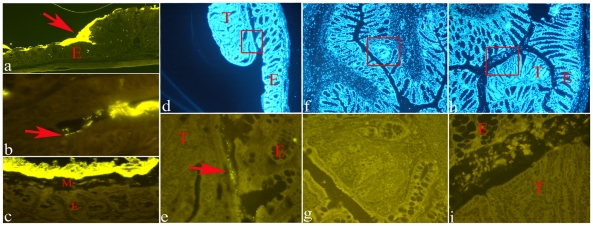
Figure 2. Representative examples of histological sections of rat colon processed with FISH (Cy3-conjugated EUB338 probe).
Panel a: normal proximal colon showing a direct contact between bacteria (bright yellow signal) and the intestinal epithelium (E) (original magnification 4×). Panel b: bacteria inside a crypt in the proximal colon (original magnification 100×). Panel c: normal distal colon showing bacteria (bright yellow signal) separated from the epithelium (E) by a layer of mucous (M) (original magnification 40×). Panel d: section of a colonic tumor (T) and its adjacent normal mucosa (E) stained with DAPI (original magnification 4×); the boxed region is shown enlarged in panel e. Panel e: presence of bacteria (arrow) at the interface between tumor (T) and normal mucosa (E) (original magnification 40×). Panel f: section of an unopened colon containing an MDF (boxed) stained with DAPI (original magnification 4×); the boxed region is shown enlarged in panel g. Panel g: no bacteria are present in the MDF (original magnification 40×). Panel h: section of an unopened colon containing a tumor (T) stained with DAPI (original magnification 4×); the boxed region is shown enlarged in panel i. Panel i: no bacteria are present in the tumor (original magnification 40×).

