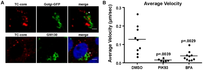
Figure 8. Localization of TC-core and markers of the TGN.
A. Huh-7.5 cells were electroporated with TC-core RNA and at 72 hours post infection cells were stained with ReAsH dye. Top panel: Cells were transduced with Golgi-GFP at 48 hours post electroporation then stained with ReAsH. Bottom panels: Cells were stained with ReAsH dye at 72 hours post electroporation and processed for immunofluorescence using an antibody directed against GM130. Arrows point to TC-core puncta colocalized with TGN markers. Scale bar is 10 µm. B. TC-core electroporated cells were incubated with DMSO, PIK93 (0.5 µM), or brefeldin A (BFA) (1 µg/ml) for 2 hours prior to ReAsh staining. Drugs were present in staining and imaging medias. Live cell imaging was performed and TC-core puncta were tracked. The average velocities were measured and are plotted. PIK93 or BFA treatment significantly reduced average velocity (unpaired Student's t-test).

